So my research involves examining diatoms, which are free-floating aquatic phytoplankton with silicified cell walls (a substance similar to glass!). A lot of my time in the lab is spent culturing different species of diatoms, which is interesting but sometimes dissatisfying–since diatoms are microscopic, most of my cultures look like vials of clear or brownish water.

Some of my small cultures chilling (literally) in a 12 degree Celsius incubator.
However, I’m always amazed when I actually look at my cultures under a microscope. Diatoms are one of the most diverse groups of phytoplankton, with over 200 known genera and an estimated +100,000 species floating around in our aquatic systems. This diversity is apparent when you magnify them, and to demonstrate that I thought I’d share some photos I took of my diatoms using a camera attached to the lab microscope. Happy Photography Friday!
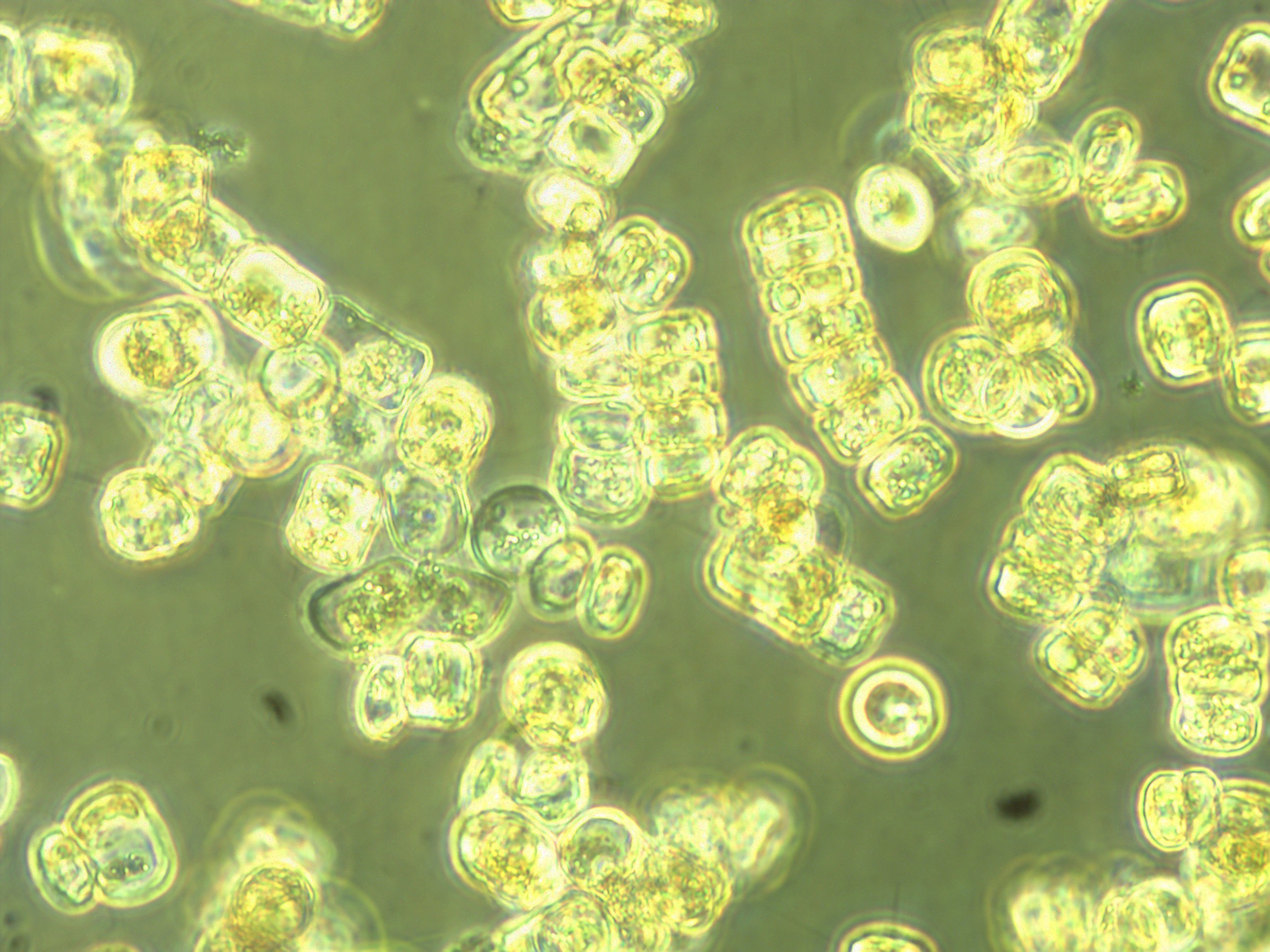
Thalassiosira sp.–a large centric diatom.

Chaetoceros sp.–another centric diatom, but this genera has long spines (called setae) growing out of each cell. These spines connect individual Chaetoceros cells into long chains.
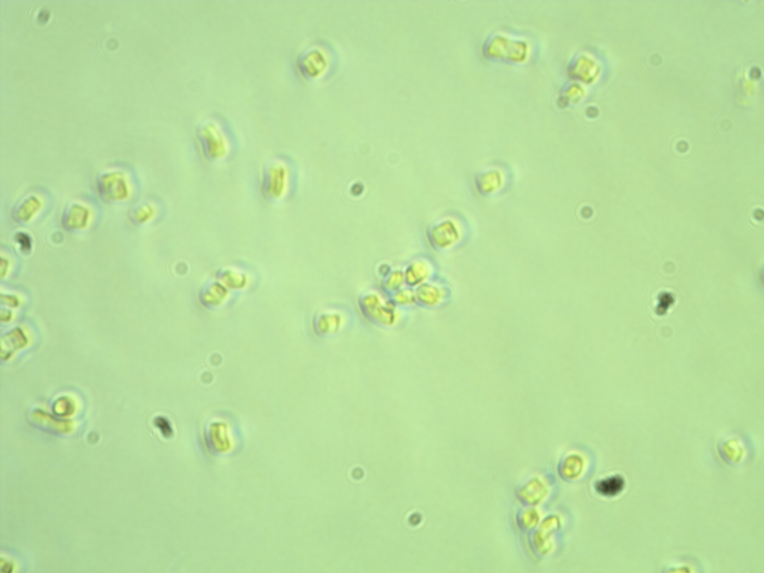
Fragilariopsis cylindrus, a polar pennate diatom. This particular culture has been lab-cultured for a long time and so the cells are much smaller than some of my other cultures. This picture was taken at a 40x, rather than 20x magnification on the microscope.

Skeletonema sp.–another centric diatom that forms long chains.
